2017
Various effects of ion implantation—Corrosion resistance of silicon carbide (SiC)-rich nanolayers produced by implantation and damage caused by radiofrequency sputtering of niobium oxide have been studied in collaboration with Institute of Technical Physics and Materials Science, Centre for Energy Research.
To produce SiC protective coatings C/Si/C/Si/C multilayer structures with different thicknesses on silicon substrates were irradiated by Ar+ and Xe+ ions at room temperature and at a fluence range of 0.5–3x10¹⁶ ion/cm2. The C, Si and SiC distributions formed by the effect of ion beam mixing were determined by Auger electron spectroscopy. In corrosion point of view the best SiC-rich layer was produced from multilayer with thicknesses of 10 nm C and 20 nm Si by implanting with 120 keV Xe with a fluence of 3x10¹⁶ Xe/cm2. As a result of the potentiodynamic experiments, the measured chemical corrosion resistance for this sample was orders of magnitude better than that of pure silicon.
To investigate the damage caused by radiofrequency sputtering, Nb2O5 layers were deposited to c-Si substrates in a gas mixture of O2 and Ar at a pressure of 0.3 Pa, at an oxygen flow of 6 ml/min and at various DC sputtering voltage selected from the range of 1.0–2.0 kV. During the rf-sputtering the energetic O+, Ar+ ions and neutral atoms may penetrate into the c-Si substrate to a depth of several nm causing displacement of host silicon atoms and consequently producing damaged regions.
In cross-sectional transmission electron microscopy image of a Nb2O5 sample with a deposition time of 100 s at 2.0-kV (not shown) a 2.9 nm thick amorphous Nb-oxide film, a 4.5 nm thick amorphous layer, which can be either amorphous SiO2 and/or amorphous Si, as well as single crystalline substrate can be observed. The partially damaged silicon layer was identified on the substrate both by Rutherford backscattering spectrometry combined with channelling (as shown in Figure 1) and by multiple angle of incidence spectroscopic ellipsometry.
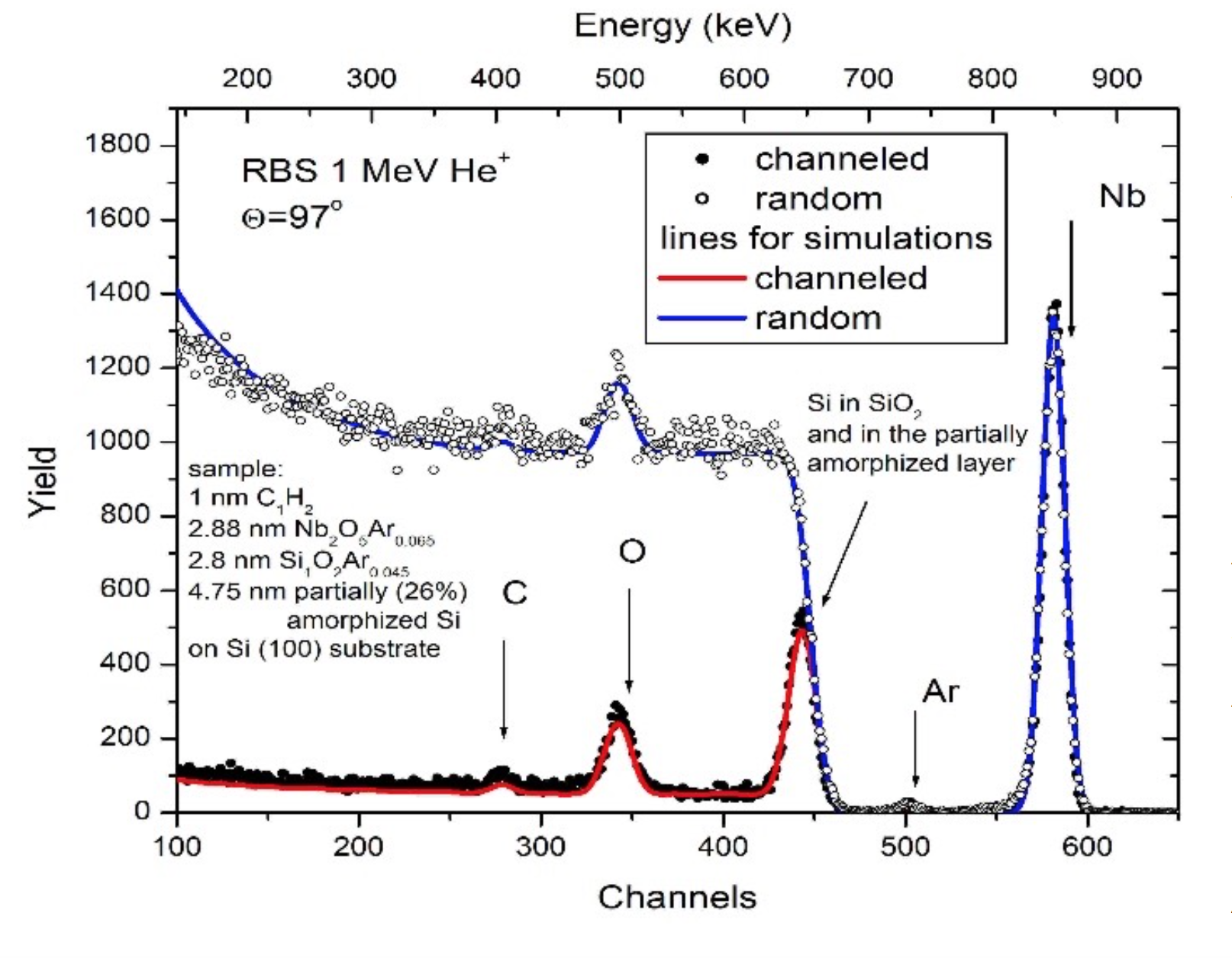
Figure 1. Channelled and random spectra taken on Nb2O5 film deposited by rf sputtering on single crystalline silicon substrate with a deposition time of 100 s at 2.0-kV.
Advanced cultural heritage research. — External milli beam PIXE was applied to determine the composition of mineral pigments from ceramics objects excavated by Romanian archaeologists from archaeological sites of important commercial settlements on Danube at the border between Ottoman Empire (Dobroudja) and Romanian Principalities – Piua Petrii and Dinogetia-Garvan.. The mineral pigments used for Turkish Miletus (late XVth-XVIth centuries) and Iznik (XVIth-XVIIth centuries) ceramics are very important for the understanding of commercial routes of late Mediaeval period. The elemental composition of glaze, green, yellow, brown and especially blue pigments was determined. The most interesting case is the one of the Co-based blue pigments (as shown in Figure 2). The origin of the raw material for these pigments in the XVIIth Century could be the mining district of Schneeberg in Germany, characterized by the presence of smaltite and erythrite minerals and this study revealed a possible trade route connection between Saxony and Ottoman Empire during those times. Arsenic is of a special importance in the case of Co blue pigments, since it was observed to appear mostly in samples dated after the year 1520.
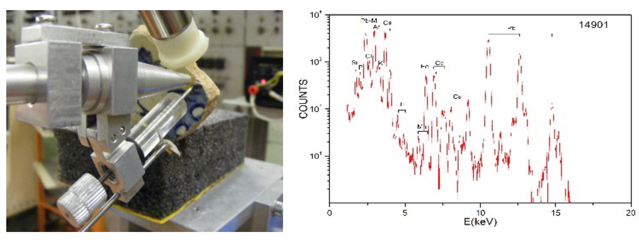
Figure 2. PIXE analysis of a dark blue area on Iznik glazed ceramics sherd (sample: CER 9)