2021
Diamond color centers. — Due to their superb light emission properties and biocompatibility, color center containing diamond nanocyrstals are promising objects for nanobiology and nanomedicine. These unique structures could be used in a marking and labelling applications, and they are pretended to replace fluorescent dyes which are suffering from photobleaching and photoionization as well as from incompatibility with living organism in some cases. Since the light absorption of tissues and different biological specimens is weaker in the first optical window (600-900 nm), thus highly fluorescent nanodiamond with emission lines lying in this wavelength range are preferred to allow efficient detection of the useful optical signal. Despite of numerous scientific reports, the efficient creation of highly fluorescent nanodiamond crystals of good quality is still challenging. We recently developed an advanced geometry utilizing plasma immersion of impurity source that could significantly increase the color center formation during the MW CVD diamond deposition process. Moreover, our technique allows to use impurity sources, required for desired color center, in solid phase and keep away from laboratory the hazardous gases, commonly applied for this purposes. On the example of silicon vacancy center, we proved that our advanced geometry appropriate to create high quality, SiV center containing nanocrystalline diamond thin films and separated nanocrystals on the silicon substrate with more than 7 times higher zero-phonon line peak intensity as it is possible for impurity source-free geometry.
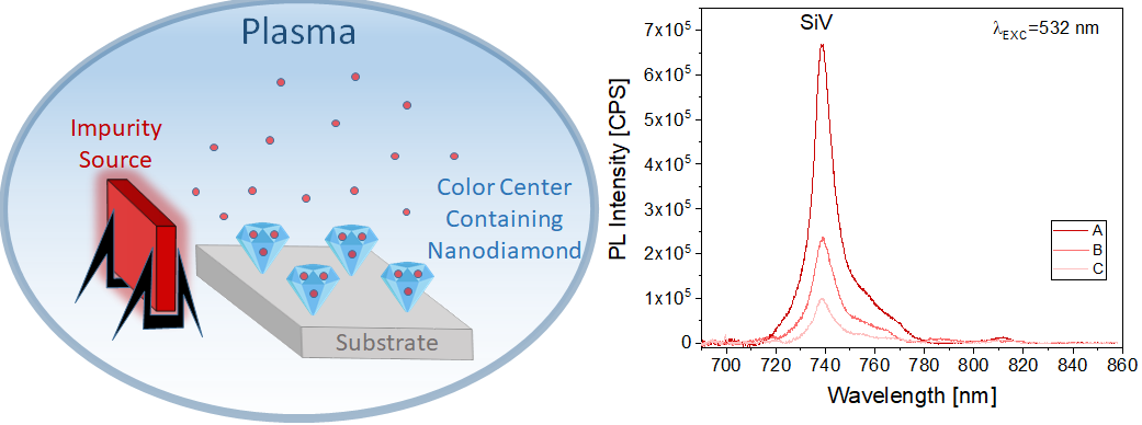
Figure 1. Schematic illustration of the plasma immersed solid impurity assisted growth of the color center containing diamond nanostructures (left). Photoluminescence (PL) spectra of the SiV center containing nanocrystalline diamond films deposited at different conditions (right): A – advanced geometry when an additional Si impurity source is immersed into the CVD plasma; B – a Si impurity source of the same dimensions placed next to the substrate; C – configuration without additional Si impurity source. The PL spectra were recorded at the same conditions using 5 mW incident power of a 532 nm diode pumped solid state laser.
Porous silicon-based nanoparticles for surface enhanced Raman spectroscopy. — Surface-enhanced Raman spectroscopy (SERS) is an emerging vibration spectroscopy technique nowadays. The key to its high sensitivity is the fabrication of metallic nanoparticle-based plasmonic structures capable of efficient amplification of the Raman signal. Porous Si (PSi) nanoparticles (NPs) were prepared by magnesiothermic reduction of the synthesized SiO2 spheres, and Ag NPs were deposited on the PSi with formaldehyde or HF (Fig.2a) [1]. In addition, silver NPs were deposited on the porous Si obtained by milling and etching the Si wafer. Silver NPs on SiO2 spheres and particles with Ag core and mesoporous SiO2 shell were also synthesized (Fig.2b). SEM analysis showed the Ag NPs size distributions between 30.1 and 64.3 nm. The synthesized samples were quite stable in aqueous media and had zeta potential between −11.5 and −44.3 mV. The samples exhibited reversible physisorption isotherms of type II except for the sample with Ag core and SiO2 (Ag–S) coating, which exhibited a type IVa isotherm with the hysteresis characteristic of mesoporous adsorbents. The BET surface areas of the samples ranged from 9 to 56 m2g-1, with the mesoporous Ag–S sample having the largest surface area of 378 m2g-1. The surface enhanced Raman spectroscopy (SERS) performance of the synthesized samples was studied by analysing the detection limit of 4-mercaptophenylboronic acid (4-MPBA) as a probe molecule at 532 nm laser excitation. The lowest limit of detection of 1×10⁻⁵ mol dm−3 was obtained with the sample in which silver NPs were deposited on silica spheres (S–Ag) (Fig.2c).
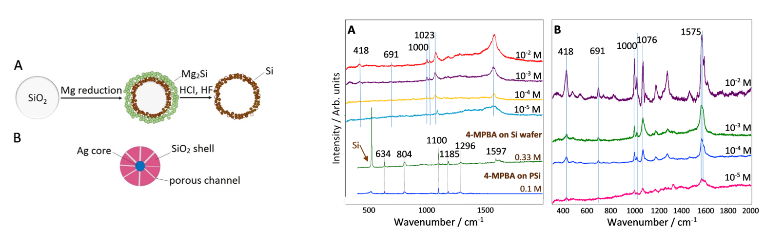
Figure 2. Schematic illustration of a) porous Si formation from synthetic SiO2, b) NP with Ag core and SiO2 coating, and c) SERS spectra of different 4-MPBA concentrations on Ag NPs on the surface of SiO2 spheres (S-Ag).
Confocal Raman micro-spectroscopy of porcine tissues with optical clearing. — Confocal Raman micro-spectroscopy (CRM) combined with optical clearing (OC) technique was used for in-depth study of the collagen in ex-vivo porcine skin in the Raman fingerprint region. The OC technique and CRM allowed one to preserve the high probing depth, signal-to-noise ratio and spectral resolution simultaneously. It was shown that the principal Raman peak intensities of skin are significantly increased at all observed depths after applied glycerol treatment for 30 min and 60 min as an optical clearing agent (Fig.3).[2]
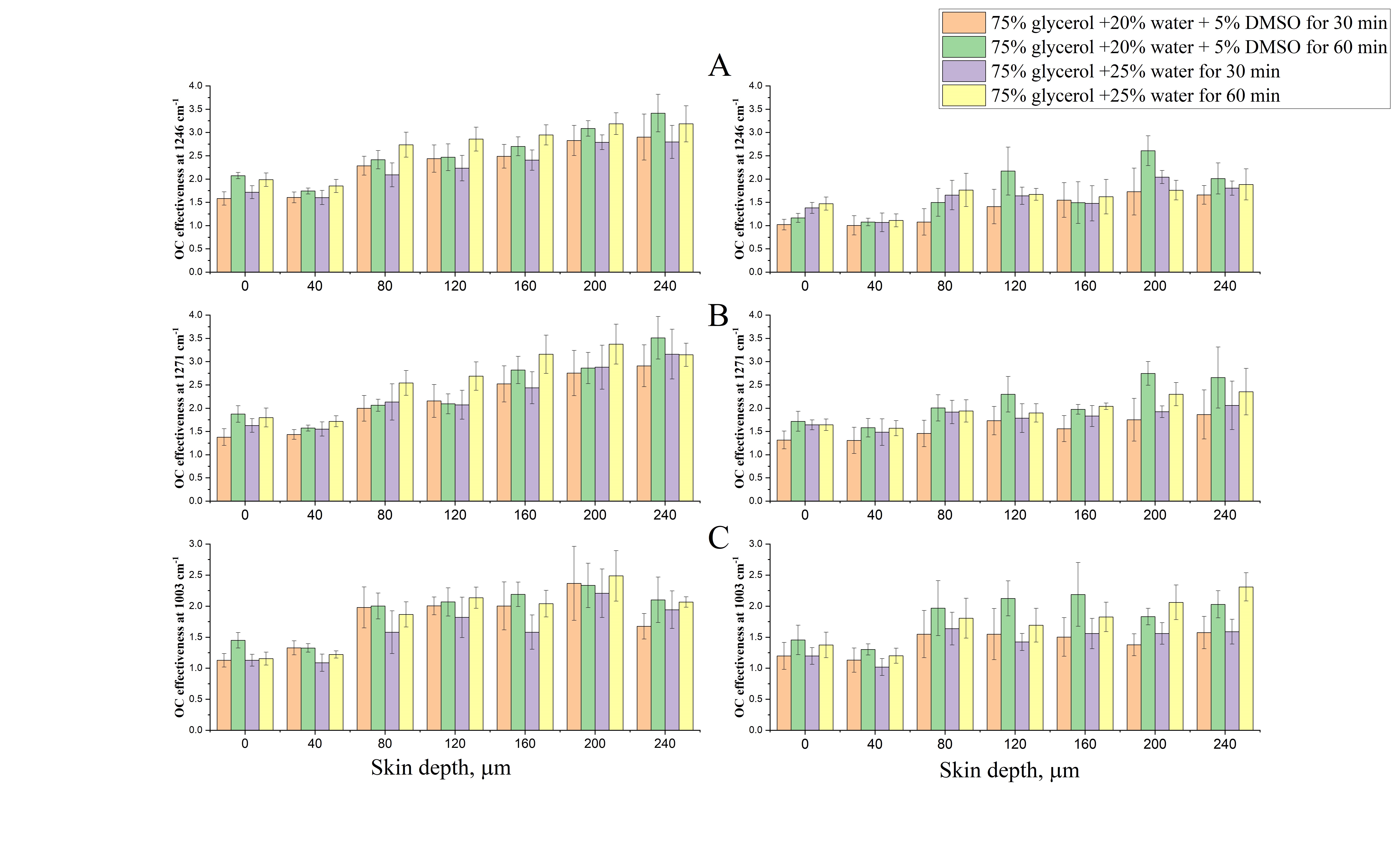
Figure 3. Optical clearing effectiveness for the porcine skin treated by different OCAs for different exposure times of 30 min and 60 min for the Raman peaks at (a) 1246 cm-1, (b) 1271 cm-1 (amide-III region) and (c) 1003 cm-1 (phenylalanine/urea) recorded with 785 nm (left) and 633 nm (right) excitation.
The influence tissue optical clearing on ex vivo confocal Raman microspectroscopy (CRM) was assessed also in porcine dura mater in order by to increase the in-depth probing of the collagen. Raman intensities were significantly increased at the depth of 250 μm after treatment with glycerol as the OC agent. The results showed that the OC can be divided into three main steps. The first one is a fast process of tissue dehydration accompanied by collagen shrinkage while the second relatively slow process is related to the glycerol penetration into the interfibrillar space of collagen combined with swelling of tissue. The third step is collagen dissociation caused by the high concentration of glycerol (Fig.4).[3]
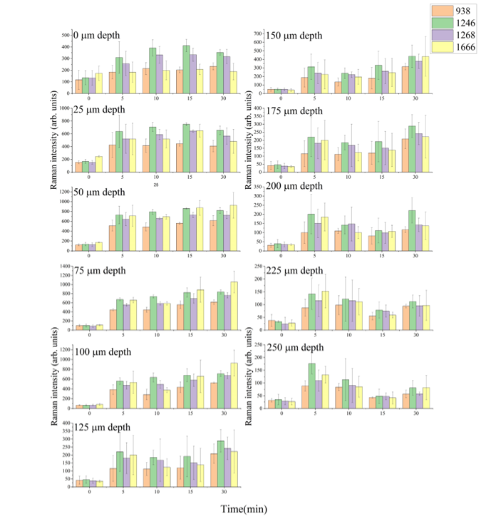
Figure 4. Evolution of the Raman bands of dura mater at 938, 1246, 1268, and 1666 cm-1 with depth between 0 and 250 μm with time, after the application of 99.0% glycerol as optical clearing agent.
Raman spectroscopic study of the degree of conversion during gamma-radiation initiated polymerization. — Diethylene glycol dimethacrylate (DEGDMA) is a biocompatible polymer forming highly crosslinked structure that is widely used in dentistry and high-pressure liquid chromatography columns. Gamma radiation-initiated polymerization is a simple and efficient technique to prepare DEGDMA polymers of different size and shape, requiring monomer mixture containing the solvent and the monomer only. In addition, bulk structures ranging from non-porous to macroporous character can be obtained changing only the type and the amount of the solvent. The degree of conversion of diethylene glycol dimethacrylate monomer in different solvents upon gamma irradiation with different doses have been studied by Raman spectroscopy and mass difference measurements (Fig.5). It has been shown that the Raman method allows determining the conversion rate of monoliths by a non-destructive and, in terms of realization, simple manner. A good agreement was found in the degree of conversion determined by Raman spectroscopy and mass difference-based techniques. The data were fitted with the Avrami equation and the observed differences in the reaction rate were explained by the different character of the DEGDMA polymerization mechanism in alcohols and other types of solvent, caused by their solubility values. It was found that the degree of conversion for DEGDMA reaches a plateau by increasing the irradiation doses and it exceeds 90% above 12 kGy, independently of the applied solvents. Raman measurements revealed also that there are DEGDMA monomers entrapped intact into the polymer matrix and/or attached to the frame only through the methacrylate group [4].
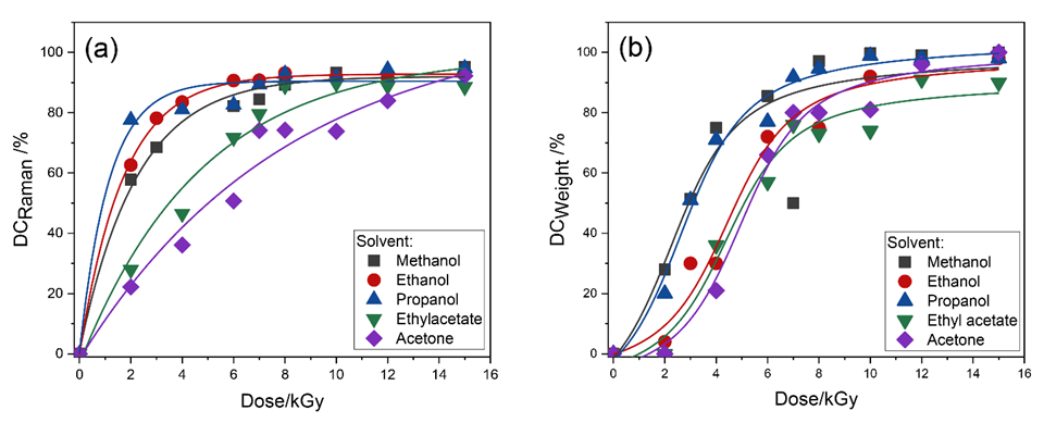
Figure 5. Degree of conversion of different DEGDMA/solvent systems with applied dose determined from (a) Raman peak intensity ratios and (b) weight difference measurements. The solid lines are curves of the Avrami fitting.